

Detect more with contrast enhanced 2D mammography and tomosynthesis fusion
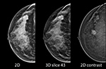
This image set from an iodine contrast mammography study was acquired under a single breast compression. The lesion pointed to with the straight arrow can be identified with the 2D image (left), although the tomosynthesis slice (center) shows the distortion associated with the lesion more clearly, and there is also iodine contrast uptake in the lesion in the contrast image (right). The lesion pointed to with the curved arrow has strong iodine uptake, but is not easily identified in the dense areas of the non-contrast mammographic image.
Using an iodinated contrast agent, I-View
