

Fujifilm’s Synapse 3D provides clinical decision support, speeds up workflow during COVID-19 emergency at Pisa University Hospital
Pisa University Hospital has received support from Fujifilm Italia to improve and speed up the analysis of lung CT images which are important for the diagnosis of COVID-19. This has been possible thanks to an update by Fujifilm of the configuration of the Synapse 3D software used by emergency room radiologists.
The hospital says this new technological development has been useful to improving management of patients affected by COVID-19, during the emergency.
Synapse3D is a 3D medical image analysis system that uses Fujifilm’s image recognition technology to construct and analyze highprecision 3D images, compiled from tomographic images from CT and MRI. It delivers 3D visualization of medical images and has applications in image-based diagnosis and surgery simulation.
Synapse 3D delivers clinical value through fast, accurate, efficient and robust image processing for radiology, cardiology and surgical preoperation simulation.
It was developed in Japan (where it is named Synapse VINCENT) and it consists of more than 50 different processing modules.
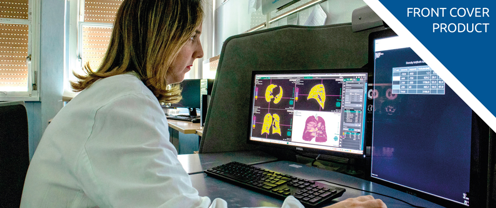
The Synapse3D Lung Analysis/Airway module enables the analysis of density ranges in the lung in a quick, easy, objective and reproducible way.
At Pisa University Hospital the configuration of Synapse 3D was completed quickly, with the study of density ranges and a feasibility analysis carried out in a few days, meaning that this updated integrated workflow has been possible to use in the hospital since April 1.
The Pisa University Hospital had already set up three levels of assessing Coronavirus patients, and with the support of Synapse 3D software, doctors have had the ability to quickly and easily identify the stage of pneumonia, and therefore hospitalise patients accordingly:
- normal hospitalisation for patients with mild pneumonia not requiring respiratory support
- assessment by a pulmonologist or intensive care doctor for patients with moderate pneumonia to plan suitable respiratory support
- intensive care assessment for patients with severe pneumonia with a view to transferring them to the intensive care unit
The density analysis provided by Synapse 3D has made it is possible to analyse the lung according to the different pixel densities of the CT images. Three groups were therefore defined based on different density ranges that allow the radiologist to evaluate the percentage of lung with lower density (emphysema), higher density (interstitial effort) and normal lung.
Dr. Chiara Romei, MD, PhD, Radiologist at the Pisa University Hospital, explained: “In a time of emergency such as this, it is crucial for us radiologists to detect accurate data as quickly as possible. Synapse 3D has enabled a quantitative analysis that is much faster and more objective than the visual analysis of the radiologist; in a couple of minutes it is possible to obtain data relating to the percentage of lung with greater and lesser density and to have a precise and objective, reproducible and shareable value.”
It is important to note that the data obtained by Synapse 3D does not replace the molecular diagnoses made through the nasopharyngeal swab (RT-PCR) and does not replace the analysis and diagnostic work by the radiologist, but instead it supports the reporting of daily exams to monitor and study the evolution of the disease, thus optimising workflow.
For medical professionals – download the take-away: https://synapse.fujifilm.eu/fujifilm-takeaway/
